


Authorized Blue-Ray Biotech Supplier
Category: Live Cell Imaging Systems
EzScope 101 Live Cell Imaging System | In-Incubator Time-Lapse Microscope
4.9 out of 5 from 78 reviews
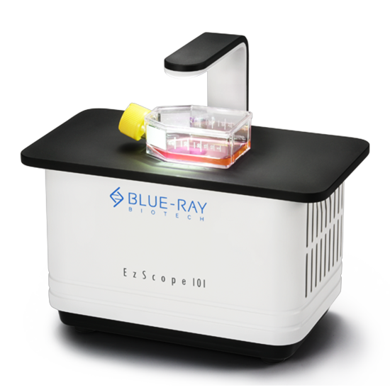
The Blue-Ray EzScope 101 is a compact live cell imaging system that sits inside a CO₂ incubator and captures brightfield time-lapse images of up to four samples, 24/7, with motorized focus, 10x/20x objectives and remote monitoring.
As a dedicated microscope in incubator, the EzScope 101 delivers continuous cell culture monitoring without disturbing the CO₂, humidity and temperature your cells depend on. It is built for live cell imaging workflows in cancer, stem cell, organoid and 3D culture research.
Interchangeable 10x and 20x objectives, a precise motorized focus and the Windows based EzCapture software produce clean time-lapse video and enable remote live cell monitoring from your workstation. Global Bioscience Solutions supplies, installs and supports the EzScope 101 across India for university core facilities, research institutes, biotech companies and pharma R&D labs.
- In-Incubator Setup
- Remote Monitoring
- Automated Time-Lapse
In-Incubator
Compatibility
Compatibility
10x / 20x
Objectives
Objectives
Motorized
Focus System
Focus System
Time-Lapse
Video Output
Video Output
Remote
Monitoring
Monitoring
Technical Specifications of the EzScope 101 Live Cell Imaging System
| System and Performance Parameters | |
|---|---|
| Brand / Model | Blue-Ray Biotech EzScope 101 |
| System Type | Automated live cell imaging system |
| Operating Environment | Inside CO₂ incubator |
| Imaging Mode | Brightfield imaging with flatfield correction |
| Objective Lenses | Interchangeable 10x and 20x |
| Focus Mechanism | Motorized focus |
| Sample Capacity | Up to four samples simultaneously |
| Software Interface | EzCapture (Windows based) |
| Data Capabilities | Measurement and analysis, spatial calibration, time-lapse output |
| Remote Access | Remote monitoring of confluence and experiment status |
Key Features of the EzScope 101 In-Incubator Microscope
- True Uninterrupted MonitoringRun live cell imaging without removing cells from the incubator. Temperature and pH shocks are eliminated, so cells behave naturally across the full experiment.
- Advanced EzCapture SoftwareProgram automated captures, apply spatial calibration, run measurement and analysis, and compile frames into time-lapse videos with the Windows based live cell imaging software.
- Multi-Sample ImagingImage up to four samples at once and compare treatments, conditions or organoid models side by side in a single run.
- Remote Access and ViewingCheck confluence and progress from your desk. Incubator imaging system remote monitoring cuts lab foot traffic and contamination risk.
- Compact, Space-Saving DesignA compact live cell microscope that frees bench space and avoids the cost of a standalone live cell imaging chamber.
Applications of Live Cell Imaging in Research and Diagnostics
- Cell Migration, Wound Healing and Proliferation AssaysRun a continuous live cell imaging wound healing assay (scratch assay), track cell migration, cell invasion and cell confluence without user intervention.
- 3D Cultures, Spheroids and OrganoidsAn ideal imaging system for the organoid lab. Monitor 3D cell culture, tumorspheroid assays and long-term organoid development.
- Stem Cell and Cardiac ResearchDocument stem cell differentiation and record cardiac cell beating events in real time.
- Cancer Biology and Drug Response StudiesFollow growth, morphology and treatment response over hours or days with automated time-lapse microscopy.
How Live Cell Time-Lapse Imaging Works with the EzScope 101
- Place the EzScope 101 inside your CO₂ incubator and connect it to the Windows PC running EzCapture.
- Load your culture vessels (up to four samples) and select the 10x or 20x objective.
- Set the capture schedule in EzCapture, then apply spatial calibration and focus.
- Monitor remotely as images are acquired automatically around the clock.
- Analyse and export measurements and time-lapse video for publication or lab meetings.
Who Uses the EzScope 101?
Cancer biology labs, stem cell and regenerative medicine groups, organoid and 3D culture facilities, university core facilities, pharma and biotech R&D teams, and diagnostic research laboratories use the EzScope 101 for reproducible, hands-off live cell imaging.
Explore Related Products and Resources
Frequently Asked Questions About the EzScope 101
What is an in-incubator live cell imaging system?
It is a compact live cell microscope that operates directly inside a CO₂ incubator. It images cells continuously without removing them from their growth conditions, which removes temperature and pH shock and lowers contamination risk.
What kind of imaging does the EzScope 101 provide?
It provides brightfield live cell imaging with flatfield correction, motorized focus and interchangeable 10x and 20x objectives, and generates time-lapse video output.
Can I monitor multiple samples simultaneously?
Yes. The EzScope 101 images up to four samples simultaneously, which suits a cell invasion assay or comparisons of organoid based disease models.
Does the EzScope 101 support remote monitoring?
Yes. The Windows based EzCapture software lets researchers check cell confluence and experiment status remotely from outside the laboratory.
What applications is the EzScope 101 used for?
It is used for wound healing assays, cell migration and proliferation tracking, confluence studies, stem cell differentiation, cardiac cell beating, 3D cell culture, tumorspheroid assays and organoid development.
What is the price of the EzScope 101 live cell imaging system in India?
Global Bioscience Solutions provides a GST quotation on request. Submit the quotation form or message the sales team on WhatsApp at +91 8310917234 for pricing, installation and support.