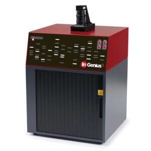
Gel Documentation System – High-Resolution Imaging for Gel Electrophoresis

A Gel Documentation System is an advanced imaging solution for visualizing, capturing, and analyzing DNA, RNA, and protein gels. It is widely used in molecular biology, life sciences, and biomedical research laboratories.
Equipped with high-resolution cameras, UV transilluminators, and fluorescence/chemiluminescence imaging capabilities, this system ensures accurate gel documentation and quantitative analysis.
Key Features of Gel Documentation System
1. High-Resolution Imaging for Accurate Documentation
- High-quality CCD or CMOS camera for sharp, detailed gel images.
- Options for fluorescence, chemiluminescence, and UV gel imaging.
2. Multiple Light Sources for Versatile Applications
- UV transilluminator for ethidium bromide-stained agarose gels.
- White light illumination for Coomassie-stained protein gels.
- Blue LED excitation for SYBR Green and other fluorophores.
3. Automated Gel Documentation and Analysis
- Easy-to-use software for image acquisition, enhancement, and quantification.
- Automated lane and band detection for protein and DNA gel analysis.
4. Compact, User-Friendly Design
- Benchtop gel doc system with touchscreen or PC connectivity.
- Ergonomic, space-saving design for small laboratories.
5. UV-Safe and Chemiluminescence Imaging
- UV shielding for operator safety during gel imaging.
- Optimized for Western blot imaging using chemiluminescent substrates.
Technical Specifications of Gel Document System
| Specification | Details |
|---|---|
| Camera Resolution | 5MP to 12MP CCD/CMOS Camera |
| Light Sources | UV, White Light, Blue LED, and Chemiluminescence |
| Transilluminator | UV (302nm, 365nm) and Blue Light |
| Imaging Capabilities | DNA/RNA Gels, Protein Gels, Western Blots |
| Software | Automated Gel Analysis, Band Detection, Image Enhancement |
| Display | Touchscreen or PC-Based Software |
| Connectivity | USB, Ethernet |
| Dimensions | Compact Benchtop Design |
| Power Supply | 110V/220V, 50/60Hz |
| Warranty | 1-2 Years |
Applications of Gel Document System
1. DNA and RNA Gel Documentation
- Imaging of agarose and polyacrylamide gels.
- Quantitative analysis of nucleic acids stained with EtBr, SYBR Green, and GelRed.
2. Protein Gel and Western Blot Imaging
- Coomassie blue, silver stain, and fluorescent-stained gels.
- Western blot analysis using chemiluminescence imaging.
3. Fluorescence and Chemiluminescence Imaging
- Detection of low-abundance proteins and nucleic acids.
- Multiplex fluorescence gel analysis.
4. Research and Diagnostics
- Used in genomics, proteomics, forensic science, and clinical diagnostics.
Why Choose the Gel Documentation System
Unmatched Imaging Precision
- High-resolution camera ensures clear and detailed electrophoresis band visualization.
- Detects faint signals in DNA, RNA, and protein gel analysis.
Versatile & Multi-Application System
- Supports multiple staining methods and gel electrophoresis techniques.
- Ideal for molecular biology, forensic science, and clinical diagnostics.
Automated Workflow for Increased Efficiency
- Auto-focus and auto-exposure reduce manual errors.
- User-friendly software simplifies data analysis and reporting.
Safe for Users and Samples
- Non-UV blue light imaging reduces radiation exposure risks.
- Ensures safe sample handling and documentation.
Cost-Effective & Low Maintenance
- Durable LED illumination reduces energy consumption.
- No need for additional UV shields or transilluminators, lowering operational costs.


